Archives of Anatomy and Physiology
Unusual Pancreatico-Mesenteric Vasculature: A Clinical Insight
Shikha Singh, Jasbir Kaur, Jyoti Arora*, Renu Baliyan Jeph, Vandana Mehta and Rajesh Kumar Suri
Cite this as
Singh S, Kaur J, Arora J, Jeph RB, Mehta V, et al (2016) Unusual Pancreatico-Mesenteric Vasculature: A Clinical Insight. Arch Anat Physiol 1(1): 001-003. DOI: 10.17352/aap.000001Background: Awareness about the variable vascular anatomy of superior mesenteric artery is imperative for appropriate clinical management. Present study not only augments anatomical literature pertaining to mesenteric vasculature but also adds to the clinical acumen of medical practitioners in their clinical endeavors.
Case summary: The present study reports the occurrence of anomalous branch, termed as accessory inferior pancreatic artery stemming from superior mesenteric artery. Additionally inferior pancreaticoduodenal artery was seen to be dividing into right and left branches instead of usual anterior and posterior branches. Right branch terminated by anastomosing with anterior branch of superior pancreaticoduodenal artery whereas left branch supplied the proximal part of the jejunum. Anomalous course of anterior branch of superior pancreaticoduodenal artery was also seen.
Conclusion: Knowledge of anomalous and variable branching pattern of superior mesenteric artery is crucial while performing various surgical and radiological procedures as it helps in minimising the intraoperative as well as postoperative complicationsCase summary: The present study reports the occurrence of anomalous branch, termed as accessory inferior pancreatic artery stemming from superior mesenteric artery. Additionally inferior pancreaticoduodenal artery was seen to be dividing into right and left branches instead of usual anterior and posterior branches. Right branch terminated by anastomosing with anterior branch of superior pancreaticoduodenal artery whereas left branch supplied the proximal part of the jejunum. Anomalous course of anterior branch of superior pancreaticoduodenal artery was also seen.
Conclusion: Knowledge of anomalous and variable branching pattern of superior mesenteric artery is crucial while performing various surgical and radiological procedures as it helps in minimising the intraoperative as well as postoperative complications.
Introduction
Eminent knowledge of morphology and embryology concerning the anomalous branching pattern of superior mesenteric artery is crucial for the successful accomplishment of various surgical, oncological and interventional procedures.
Superior mesenteric artery (SMA) stems as one of the ventral branches of abdominal aorta, behind the body of pancreas 1 cm below the origin of coeliac trunk (L1/2 intervertebral disc level). It supplies the derivatives of midgut extending from the 2nd part of duodenum (at the opening of common bile duct) to the junction between right 2/3 and left 1/3 of transverse colon. SMA gives off several branches including inferior pancreatico-duodenal artery, jejunal, illeal, ileocolic, right colic and middle colic arteries [1]. Numerous variations of the SMA concerning its origin, course and branching pattern have been reported in literature [2,3]. In the present study, the authors report the occurrence of an anomalous branching pattern of SMA, its embryological basis and clinical significance.
Awareness of such anatomical variations is imperative in preventing fatal complications following important surgical procedures like pancreatic resections. Thorough anatomical knowledge of origin and branching pattern of superior mesenteric artery is also significant in invasive procedures like celiacography and chemoembolisation of pancreatic and liver tumors. The present study describes in detail a unique variation of superior mesenteric artery, which may be of utmost significance for the present day physician and surgeon in accurate interpretation of diseases with vascular involvement and optimal clinical management. The present study is a humble attempt to avoid iatrogenic injuries from surgical and interventional radiologic procedures.
Result
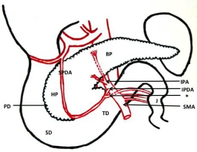
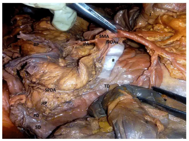
During the course of preclinical educational teaching program unusual branching pattern of superior mesenteric artery was observed in a 62-year-old Indian male cadaver. An anomalous inferior pancreatic branch was seen to be arising from superior mesenteric artery, 1.25cm distal to its origin from abdominal aorta at the level of 1st lumbar vertebra. The artery coursed superiorly towards the inferior border of pancreas and bifurcated into anterior and posterior branches, at a distance of 1cm proximal to the level of origin. The anterior branch was 0.9cm in length and entered the posterior surface of the pancreas near its inferior border. The terminal part of the branch was at a distance of 10 cm from the tail of pancreas. The posterior branch was 0.6 cm in length and coursed upwards to enter the posterior surface of the gland near its upper border, at a distance of 9cm from the tail of pancreas.
Interestingly, inferior pancreatic duodenal artery (IPDA) also exhibited an anomalous branching pattern. Emanating 2.1 cm distal to the origin of superior mesenteric artery, the IPDA was observed as a thin branch. The credence of the report lies on the observation that the IPDA divided into right and left branches instead of usual anterior and posterior inferior pancreaticoduodenal branches. The right IPDA anastomosed with anterior branch of superior pancreaticoduodenal artery. However, the left branch took an aberrant course and supplied the proximal part of jejunum. In the present study as the left branch of IPDA supplied the jejunum and right branch of IPDA supplied pancreas and duodenum, it was termed inferior pancreaticoduodenojejunal artery.
In addition, a variable course of anterior branch of Superior pancreaticoduodenal artery was observed. The artery took an anomalous course superficial to anterior surface of head of pancreas instead of its normal course in the groove between 2nd part of duodenum and head of pancreas. Crossing the anterior surface of head of pancreas, the anterior branch of Superior pancreaticoduodenal artery entered the pancreaticoduodenal groove, 1 cm distal to the junction of 2nd and 3rd part of duodenum. The terminal part of the artery ended by anastomosing with right branch of IPD (Figures 1,2).
Discussion
Superior mesenteric artery, an unpaired ventral branch of abdominal aorta commences at the level of first lumbar intervertebral disc,1 cm below the coeliac trunk. During embryogenesis, three groups of collateral arteries arise from abdominal aorta including somatic intersegmental, lateral splanchnic and ventral splanchnic. Ventral Splanchnic branches appearing initially as paired vessels are connected by ventral longitudinal anastomosis (lang anastomosis) [4]. These paired vessels coalesce in the midline forming four roots. The first and fourth root forms celiac trunk and superior mesenteric artery respectively, however the second and third roots disappear. The branches of celiac trunk and superior mesenteric artery are kept separated by Lang anastomosis. Variations occurring at these separation levels may lead to the formation of collateral branches [4]. Occurrence of these variations can also be explained by typological theory according to which the origin of celiac-mesenteric vasculature forms six pairs of left and right vessels (subphrenic, upper, middle and lower ventricular, and upper and lower intestinal arteries). These vessels amend during late phases of fetal development and during these changes persistence of parts destined to disappear or vice versa may result in formation of additional branches [5]. Some authors opined that during development, differences in midgut rotation and its physiological herniation, splenic migration to left, hemodynamic, genetic, racial, evolutionary and ontogenic changes in the abdominal viscera, might lead to such variations [6].
In the present case report, the authors observed an anomalous branch, termed as inferior pancreatic artery arising from the Superior mesenteric artery, 1.25cm distal to its origin from the abdominal aorta. The artery shortly after its commencement divided into anterior and posterior branches, both the branches entered the substance of body of pancreas. In previous studies, Inferior pancreatic artery has been observed to arise from dorsal pancreatic artery in 80% of the cases or as a continuation of left branch of anterior superior pancreaticoduodenal artery in 10% of the cases. However, it was observed to arise from Superior mesenteric artery in only 1 % of the cases [7]. Thus, according to literature, the origin of inferior pancreatic artery from Superior mesenteric artery is a rare occurrence. Such variable pancreatic vasculature becomes crucial during management of gastric and duodenal ulcers, limited pancreatic resection as well as transplantation of pancreas as it may result in superfluous bleeding.
Further, the present study also describes the variable division of Inferior pancreaticoduodenal artery into right and left branches instead of normal anterior and posterior branches. Inferior pancreaticoduodenal artery normally arises from superior mesenteric artery as its first branch and divides into anterior and posterior branches supplying the distal part of duodenum along with head and uncinate process of pancreas. However, in the present case report IPDA showed an anomalous division into right and left branches. The right branch ended by anastomosing with anterior branch of superior pancraeticoduodenal artery. The left branch was unique as it supplied the proximal part of jejunum and hence the IPDA was designated the nomenclature of inferior pancreaticoduodenojejunal artery by the authors. Possibility of such variants should be borne in mind by Surgeons, as their ligation during pancreatic resection operations would also proximal part of jejunum ischemic.
Also, it was observed that the anterior branch of superior pancreaticoduodenal artery coursed in front of anterior surface of head of pancreas instead of coursing in the groove between second part of duodenum and head of pancreas. The branch entered the pancreaticoduodenal groove 1cm distal to the junction of second and third part of duodenum and ended by anastomosing with the right branch of Inferior Pancreaticoduodenal artery. Distance of anterior pancreaticoduodenal arcade from second part of duodenum was reported to be ranging from 0 to 3cm [8]. In the present case however, the distance was observed to be 3.5cm. Both pancreatic head and duodenum share blood supply through pancraticoduodenal arcades; hence awareness about their variations is crucial during 95% pancreatic resection, because colossal interference with these arcades may result in duodenal ischemia [8].
The treatment of pancreatic ailments like chemotherapy for pancreatic carcinomas via superselective embolization [9] or arterial infusion [10], intrarterial stem cell infusion for diabetes require eminent knowledge of topography of blood vessels further enhancing the importance of studying vascular anatomy of SMA. Anatomical knowledge of such variations is imperative for surgical procedures such as liver transplantation and resection, gastrectomy, billiary reconstruction, right hemicolectomy and resection of transverse colon. Awareness of such variations is of utmost significance in surgical procedures like pancreaticoduodenectomy, aortic surgery, pancreatic and hepatobilliary malignancy, and local inflammation accompanying a billiary stent or obesity.
Variant vascular anatomy might also result in the unfortunate faulty analysis of angiograms. Identification of these aberrant vessels is therefore vital to avoid iatrogenic injuries. Awareness of variant pancreatic vasculature is essential for the treatment of early cases of pancreatic cancer as mistaken ligature of branches of pancreaticoduodenal vessel may end up damaging preserved pancreatic portion [11]. In surgical management of acute necrohemorrhagic pancreatitis, a wrong ligature may result in uncontrollable hemorrhage and necrosis of a normal part of the gland [12].
Knowledge of variant vascular anatomy of pancreaticoduodenal region is imperative before any planned surgery of the region. It provides a better insight of the region thereby increasing the success rate of the procedure and prevention of any postoperative complications.
Thus the uncommon and rare variations in the branching pattern of arteries of gut are of immense importance for present day surgeon, as injury to these vessels might result in severe hemorrhage and pre and post complications. The present case report describes the unique variations of branches of superior mesenteric artery, which may be of utmost relevance for the radiologists and gastroenterologists for appropriate clinical management.
- Standring S (2008) Gray’s anatomy – the anatomical basis of clinical practice. 40th edition, Elsevier Churchill Livingstone, Edinburgh 1130-1131. Link: https://goo.gl/BRg1mF
- Günenç C, Denk CC (2006) Combined unusual anatomical variations of the superior mesenteric and right renal arteries. Clin Anat 19: 716–717. Link: https://goo.gl/wxedkn
- YI SQ, Li J, Terayama H, Naito M, Iimura A, Itoh M (2008) A rare case of inferior mesenteric artery arising from the superior mesenteric artery, with a review of the review of the literature. Surg Radiol Anat 30: 159-165. Link: https://goo.gl/7Oa19T
- Tandler J (1904) Über die Varietäten der Arteria coeliaca und deren Entwicklung. Anat Hefte 25: 473–500. Link: https://goo.gl/KXFhkd
- Murakami T, Mabuchi M, Giuvarasteanu I, Kikuta A, Ohtusuka A (1998) Coexistence of rare arteries in the human Celiacomesenteric system. Acta Med Okayama 52:239-244. Link: https://goo.gl/f1y9ab
- Reuter SR, Redman HC (1977) Gastrointestinal angiography. 2nd Edition, WB Saunders, Philadelphia 31-65. Link: https://goo.gl/H9qGsd
- Woodburne RT, Olsen LL (1951) The arteries of the pancreas. Anat Rec 111: 255–270. Link: https://goo.gl/XqNSiJ
- Chavan NN, Wabale RN (2015) Arterial arcades of Pancreas and their variations. International J of Healthcare and Biomedical Research 3: 23-33. Link: https://goo.gl/ZaEX9O
- Lin Y, Yang X, Chen Z, Tan J, Zhong Q, et al. (2010) Demonstration of the dorsal pancreatic artery by CTA to facilitate super selective arterial infusion of stem cell into the pancreas. Eur J Radiol 81: 461-465. Link: https://goo.gl/g2Dljx
- Takamori H, Kanemitsu K, Tsuji T, Tanaka H, Chikamoto A, et al. (2005) 5–Fluorouracil intra – arterial infusion combined with systemic Gemcitabine for unresectable pancreatic cancer. Pancreas 30: 223-226. Link: https://goo.gl/hywW4u
- Loos M Kleeff J, Friess H, Büchler MW (2008) Surgical treatment of Pancreatic Cancer. Ann N Y Acad Sci 1138: 169-180. Link: https://goo.gl/j5AmzN
- Bertelli E, Di Gregorio F, Bertelli L, Mosca S (1995) The arterial blood supply of the pancreas: a review. I. The superior pancreaticoduodenal and the anterior superior pancreaticoduodenal arteries. An anatomical and radiological study. Surg Radiol Anat 17: 97-106. Link: https://goo.gl/lQ7Yh7
Article Alerts
Subscribe to our articles alerts and stay tuned.
 This work is licensed under a Creative Commons Attribution 4.0 International License.


 Save to Mendeley
