Archives of Anatomy and Physiology
Double outlet right ventricle, total anomalous venous return, total anomalous hepatic venous drainage and supra mitral ring in a child with Ivemark`s syndrome
Girish Gowda SL1*, Seetharama Bhat PS1, Jayaranganath Mahimarangaiah2 and Cholenahally NM2
2Department of cardiology, Sri Jayadeva Institute of Cardiovascular Sciences and Research, India
Cite this as
Girish Gowda SL, Seetharama Bhat PS, Mahimarangaiah J, Cholenahally NM (2017) Double outlet right ventricle, total anomalous venous return, total anomalous hepatic venous drainage and supra mitral ring in a child with Ivemark`s syndrome. Arch Anat Physiol 2(1): 018-019. DOI: 10.17352/aap.000006Double outlet right ventricle, total anomalous venous return, total anomalous hepatic venous drainage and supra mitral ring with Ivemark syndrome is an unusual combination of cardiac malformations. These complexities of congenital anomalies can pose problem in preoperative diagnosis and surgical management. We present investigation and surgical management of a three month old child with these mentioned rear combination of anomalies and a brief revive of literature.
Introduction
Double outlet right ventricle, total anomalous venous return, total anomalous hepatic venous drainage and supra mitral ring in a child with Ivemark syndrome is an unusual combination of cardiac malformations which has not been reported till now in the world literature. These complex combinations of cardiac malformations can be surgical corrected.
Case Report
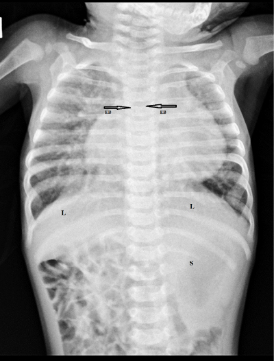
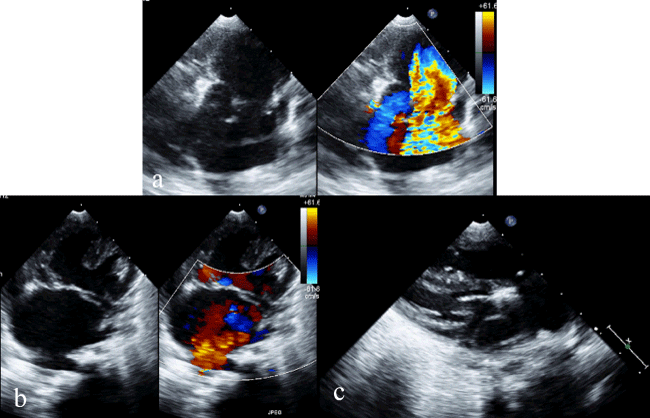
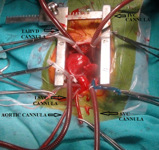
A three month old female child was referred to our institution with a history of cyanosis since birth for evaluation and treatment. There was mild cyanosis present with a grade III/IV systolic murmur along the left and right sternal border, heard more over the third left interspace, which radiated well into the second right interspace. All peripheral pulsations were well felt. Chest and abdomen x-ray showed bilateral eparterial bronchi, symmetrical transverse liver and ipselateral side of liver and stomach (Figure 1). ECHO showed findings of DORV, cardiac type of total anomalous venous return, total anomalous hepatic venous drainage in to RA and supra mitral ring with normal drainage of inferior venacava into RA (Figure 2). With the above mentioned diagnosis patient was taken for surgery. Both atrial appendages were morphologically identical to right atrium and child had LSVC. All three venacavas and anomalous hepatic vein were cannulated for cardiopulmonary bypass (Figure 3) (video). On opening the RA the diagnosis of cardiac total anomalous venous return, DORV and supra mitral ring were confirmed. Resection of supra mitral ring, rerouting of the pulmonary veins to the left atrium and intra ventricular tunneling of VSD to aorta was performed. Child was extubated on the second postoperative day.
Discussion
Ivemark’s syndrome is a rare congenital anomaly which is inherited in an autosomal recessive fashion and is estimated to occur in 1 in 10,000 to 40,000 live births [1,2]. Ivemark’s syndrome consists of situs ambiguus with intrathoracic and abdominal visceral heterotaxy. Intracardiac defects associated with Ivemark syndrome includes common atrium ventricular septal defect with a common atrioventricular valve, common atrium, interruption of the inferior vena cava with azygos venous continuation to the atrium, direct hepatic venous drainage to the atrium, DORV, bilateral superior venacava and various other cardiac anomalies [3]. With extensive searching of world literature through PubMed no case with Ivemark’s syndrome associated with supramitral ring were found. Hence this is the first reported case of this combination of anomalies. In some cases of right isomerism, right and left sided atrial origin of the P wave is present at different times of ECG reflecting the activity of bilateral sinus nodes [4]. Our case also had this mentioned ECG finding. Atrioventricular block is very uncommon in right isomerism whereas varying degrees of atrioventricular block are common in left isomerism. Patients with right isomerism may occasionally present with serious extra cardiac anomalies [5]. Our reported case had no extra cardiac anomalies. The IVC drains into the heart through azygos vein more often in patients with left atrial isomerism whereas in right atrial isomerism the hepatic veins drains into the right atrium with an interruption of the IVC or without interruption of IVC [6,7]. Cardiac anomalies are more frequent in right atrial isomerism and lead to a mortality of 80% to 95% in the first year of life without their prior surgical correction [8]. In the absence of cyanosis or cardiac anomalies, patients with asplenia are at greater risk of dying from sepsis than from congenital heart disease. Our reported case had extremely rare combination of cardiac anomalies which is first in the word literature to be reported.
- Hurwitz RC, Caskey CT (1982) Ivemark syndrome in siblings. Clin Genet 22: 7-11. Link: https://goo.gl/WZjhRP
- Cesko I, Hadjú J, Tóth T, Marton T, Papp C et al. (1997) Ivemark syndrome with asplenia in siblings. J Pediatr 130: 822-824. Link: https://goo.gl/xWBSzc
- Harold Chen (2006) Atlas of Genetic Diagnosis and Counseling: Ivemark Syndrome 549-552. Link: https://goo.gl/Ry92bV
- Wren C, Macartney FJ, Deanfield JE (1987) Cardiac rhythm in atrial isomerism. Am J Cardiol 59: 1156-1158. Link: https://goo.gl/jqqzA5
- Ticho BS, Goldstein AM, Van Praagh R (2000) Extracardiac anomalies in the heterotaxy syndromes with focus on anomalies of midline-associated structures. Am J Cardiol 85: 729-734. Link: https://goo.gl/WgNnoh
- Anderson R, Adams P, Bruke B (1961) Anomalous inferior vena cava with azygos continuation (infrahepatic interruption of inferior vena cava). J Pediatr 59: 370-383. Link: https://goo.gl/zRdZjp
- Sheley RC, Nyberg DA, Kapur R (1995) Azygous continuation of the interrupted inferior vena cava: a clue to prenatal diagnosis of the cardiosplenic syndromes. J Ultrasound Med 14: 381-387. Link: https://goo.gl/BXJ8mG
- Hashmi A, Abu-Sulaiman R, McCrindle BW, Smallhorn JF, Williams WG et al. (1998) Management and outcomes of right atrial isomerism: a 26-year experience. J AmColl Cardiol 31: 1120-1126. Link: https://goo.gl/32cJ8f
Article Alerts
Subscribe to our articles alerts and stay tuned.
 This work is licensed under a Creative Commons Attribution 4.0 International License.


 Save to Mendeley
