Archives of Anatomy and Physiology
Aortic Arch Morphometry and its clinical implication –A computed tomography study
Hema Nagpal1*, PK Sharma2, Jyoti Chopra3 and Rajni Patel4
2Professor and Head, Department of Anatomy, ERA Medical College, Lucknow, UP, India
3Professor, Department of Anatomy, King George’s Medical University, Lucknow, UP, India
4Associate professor, dept of anatomy, SIMS, HAPUR, UP, India
Cite this as
Nagpal H, Sharma PK, Chopra J, Patel R (2018) Aortic Arch Morphometry and its clinical implication –A computed tomography study. Arch Anat Physiol 3(1): 005-008. DOI: 10.17352/aap.000011Aortic arch is a challenging site for endovascular repair. In the present study morphometric details of aortic arch such as aortic arch angle, aortic arch type and angle of aortic arch curvature were observed with an objective to provide normogram of these parameters for north indian population as the literature is lacking with these values. These parameters were corelated with age and gender also. It is a pilot study of its own kind.
Introduction
Aorta is the main arterial trunk that delivers oxygenated blood from the left ventricle of the heart to the tissues of the body. The major branches of arch of aorta are the great ways for blood supply to the head and upper limb, and are of particular interest in clinical angiography. The most common branching pattern of the aortic arch in humans comprises of three great vessels; first, the brachiocephalic trunk then the left common carotid artery and finally the left subclavian artery. The aortic arch and its branches is important anatomical structure for both surgeons and interventionalists. Aneurysms and dissections of the aortic arch need to be treated with complex surgical procedures such as deep hypothermic circulatory arrest and selective antegrade cerebral perfusion. These procedures evolved to enable replacement of the aortic arch and reconstruction of its continuity to the aorta with less risk of ischemic and embolic cerebral damage. For this the knowledge of morphometric data of the aortic arch can be of help for conceiving, designing and optimizing all types of diagnostic and therapeutic interventions involving the aortic arch and its branches.
Materials and Methods
The present study was conducted on 55 patients, including 31 males and2 4females. The patients underwent computed tomographic angiographic scan for various indications, were included in the study. Age of subjects ranged from 3 months to 75 years across 5 age groups. Written informed consent from the patients was obtained. The images of patients with previous history of allergy to contrast agent, renal insufficiency,distortion of anatomy of arch of aorta due to any pathology were excluded from this study. A region of interest was drawn on the aorta at the level of the diaphragm. After an appropriate delay to allow passage of the contrast agent into the renal arterial circulation, a series of thin cuts (0.9mm) were obtained throughout the aorta. Morphometry of arch of aorta were studied in axial, multiplanar reconstructions (MPR) images, and in volume rendered images.
The parameters observed were:
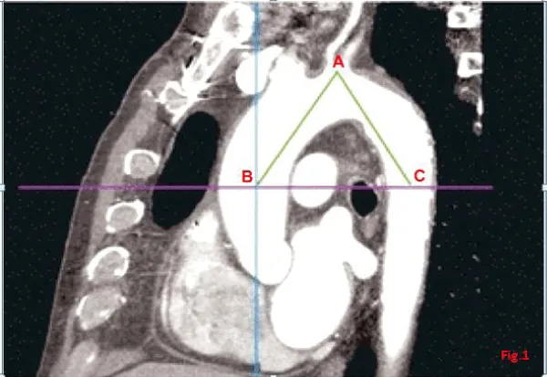
1. Aortic arch angle- It was defined as the angle between a line connecting the highest point of the aortic arch and a mid-luminar point of the ascending and descending aorta at the height of the bifurcation of the pulmonary trunk in the parasagittal plane [1] (Figure 1).
2. Aortic arch type- There was three types of arches, type I, II and III in which the vertical distance from the origin of the brachiocephalic trunk (BCT) to the top of the arch in parasaggital image was measured,if this distance was less than the diameter of LCA it was termed Type I arch,if the distance was less than double of diameter of LCA but more than its diameter this was labelled as Type II; and if the distance was more than the double of diameter of LCA it was termed Type III arch [2] (Figure 2).
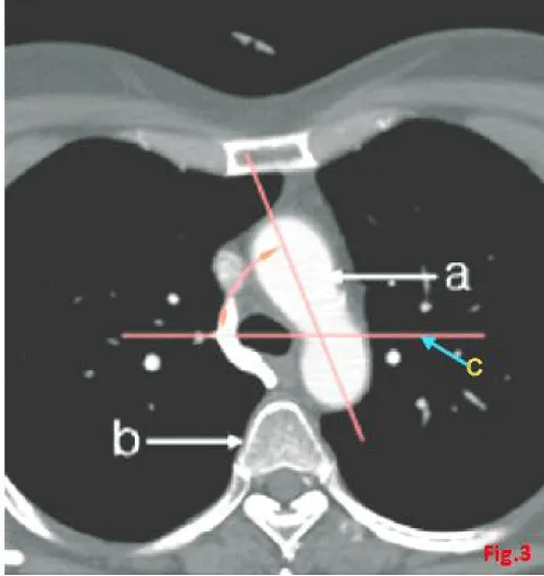
3. Angle of aortic arch curvature-It was the angle formed by aortic arch from coronal plane in axial view (Figure 3).
Results
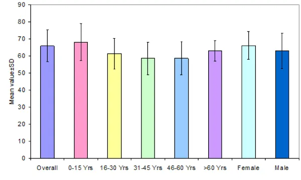
Age of subjects ranged from 3 months to 75 years across 5 age groups. Except for age groups 0-15 and 61-75 years, all the three age groups had 13 (23.64%) subjects each. In age group 0-15 years there were 10 (18.18%) and in age group 61-75 years, there were 6 (10.91%) cases. Mean age of subjects was 36.4±19.2 years. The study population consisted of 24 (43.64%) females and 31 (56.36%) males. The efforts were made to include sufficient number of subjects from each age group, Aortic arch angle were measured in parasaggital images with the help of electronic caliper. It ranged from 46.4° to 85.5o with a mean value of 65.8°±9.4o. The most common range of angle observed in the study population was 60º-70º in 47.27% (n=52) cases.
Among different age groups, the mean was found to be minimum in age group 15-30yrs (63.4±9.1o) and maximum in Groups 0-15 yrs and 46-60yrs (68.1±11.0o and 68.1±9.6o), however, the difference in different age groups was random in nature and did not account for any significant difference statistically (p=0.592). Females had higher mean value as compared to males yet the difference was not significant statistically. As age and gender wise differences were not significant, hence the normative range (63.3°-68.3°) for entire sample could be proposed as representative of all age and gender groups (Table 1,2, Figure 4) .
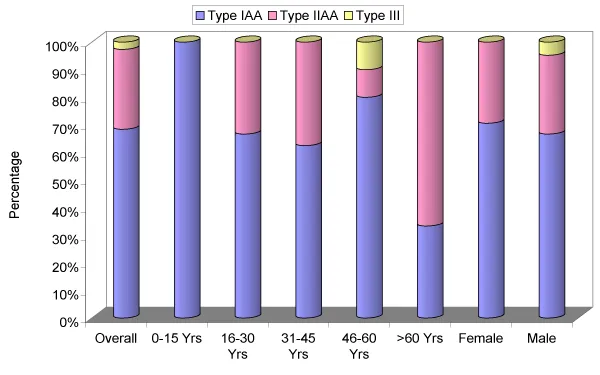
Type of aortic arch could be categorized in 38 subjects as the subjects having common trunk for BCT and LCA were excluded. Total 26(68.42%) aortic arches were detected as Type I, 11 (28.95%) as Type II and only 1 (2.63%) aortic arch was categorized as Type III. Except for age group >60 years, where Type II was more common as compared to other types, in all other age groups Type I was the most common type. Statistically there was no significant difference in type of aortic arch in different age groups and gender (p>0.05) (Table 3, Figure 5).
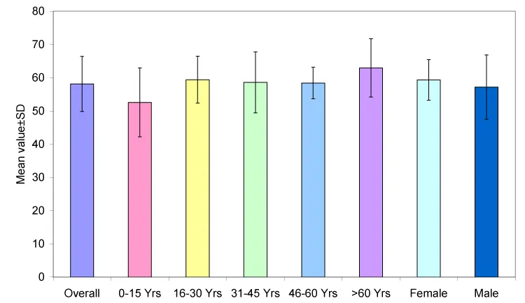
Angle of aortic arch curvature to the coronal plane was studied in 55 subjects and it was found to range from 30o to 77.6o with a mean value of 58.1o±8.3o. Among different age groups, the mean was found to be maximum in age group 61-75yrs (63.0±8.8o) and minimum in age group 0-15yrs (52.6±10.4o), however, the difference in different age groups was random in nature and did not account for a significant difference statistically (p=0.139). Gender wise, males had a higher mean value as compared to females yet the difference was not significant statistically (p=0.338). As age and gender wise differences were not significant, hence the normative range for entire sample could be proposed as representative of all age and gender groups (Table 4, Figure 6).
Discussion
Planning for endovascular interventions involving the aortic arch requires a detailed familiarity with arch and supra-aortic branch morphology. The aortic arch poses several challenges in terms of morphology, significant hemodynamic forces, and the presence of supraaortic branches. Thus data on aortic arch morphology are necessary to increase the utility of totally endovascular aortic arch repair and to pave the way for ‘‘off-the-shelf’’ fenestrated/ branched stent-grafts. Lack of knowledge can lead to relatively higher rates of complications and reinterventions following endovascular treatment of aortic arch lesions.
In the present study there was a great difference between the minimal and maximal values of aortic arch angle (46.40° and 85.50°), which points towards a significant variability in steepness of aortic arch. These values are comparable with the result of Demertzis et al. who found it between 43° -80° [3]. We also studied the effect of age and gender on aortic arch angle and found that it did not show any significant relation with age but in females it was wider than males but this difference was statistically non-significant.Aortic arch angle represents the steepness of aortic arch, so less the angle, more the steepness, resulting into more difficulty in interventional maneuvers for supra-aortic artery.
In the present study, we found type I arch to be more frequently presented (n=26, 68.42%) followed by type II (n=11, 28.95%) and only one (n=1,2.63%) aortic arch was categorized as Type III. Whereas Demertzis et al. observed Type I aortic arch in 47%, type II in 36% and type III in 17% patients [3]. Liu also found type I arch in 44.7%, type II arch in 31.4%, type III arch in 23.9% of cases [4]. The prevalence of type I arch was quite high and that of type III arch was quite low in present study. In the present study type 1 was the most common type in all age groups except for age group >60 years, where type II was more common. This finding was similar to the finding of Liu [4]. Although aortic arch type did not show any significant corelation with age and gender (p>0.05) as also observed by Demertzis et al. [3].
The aortic arch type gives an additional impression of steepness of the aortic arch [2].The type III arch was correlated to the maximum grade of difficulty in interventional maneuvers .We found type I to be most common in our study so there should be minimum difficulty of interventional maneuvers for supra-aortic artery access in our population.
In present study average angle of aortic arch curvature to coronal plane was 58.11° (range, 30°-78°) which was almost similar to the value observed by Shin et al. (62.2° with a range of 30°-90°) [5]. Angle was found to be lowest (52.57+10.38 mm) in subjects aged 0-15 years and highest (62.95-8.78 mm) in subjects aged 61-75 years, but this difference was statistically non-significant (p=0.139).Mean aortic arch curvature of female subjects was found to be higher (59.34+6.09) as compared to male subjects (57.16+9.66) but this difference was also statistically non-significant (p=0.338).
So this study provides a basic anatomical information to catheterize aortic arch and its branches for safely performing endovascular surgery.
- Agnoletti G, Ou P, Celermajer DS, Boudjemline Y, Marini D, et al. (2008) Acute angulation of the aortic arch predisposes a patient to ascending aortic dilatation and aortic regurgitation late after the arterial switch operation for transposition of the great arteries. J Thorac Cardiovasc Surg 135: 568–572. Link: https://goo.gl/2ShUJt
- Madhwal S, Rajagopal V, Bhatt DL, Bajzer CT, Whitlow P, et al. (2008) Predictors of difficult carotid stenting as determined by aortic arch angiography. J Invasive Cardiol 20: 200–204. Link: https://goo.gl/X1d96D
- Demertzis S1, Hurni S, Stalder M, Gahl B, Herrmann G (2010) Aortic arch morphometry in living humans. J Anat 217: 588–596. Link: https://goo.gl/i3vbKv
- Luis A Yeri Jorge E Gómez (2011) Sergio Fontaneto & Marcia EspósitoVariation of the Origin of Aortic Arch Branches: In Relationship with Plates of Atheroma Int. J. Morphol 29: 182-186 Link:
- Shin IY, Chung YG, Shin WH, Im SB, Hwang SC, et al. (2008) A Morphometric Study on Cadaveric Aortic Arch and its branches in 25 Korean Adults: The perspective of Endovascular surgery. J Korean Neurosurg Soc 44: 78-83. Link: https://goo.gl/fkNwqf
Article Alerts
Subscribe to our articles alerts and stay tuned.
 This work is licensed under a Creative Commons Attribution 4.0 International License.


 Save to Mendeley
