Archives of Anatomy and Physiology
The association between cervical lordosis and age, sex, history of cervical trauma and sedentarity: A CT study
David Ezra1, Leonid Kalichman2, Azaria Simonovich3, Jonathan Droujin3, Ella Been4 And Deborah Alperovitch-Najenson2*
2Department of Physical Therapy, Recanati School for Community Health Professions, Faculty of Health Sciences, Ben-Gurion University of the Negev, Beer Sheva, 8410501 Israel
3Radiology Department, Barzilai University Medical Center, Ashkelon, 7830604 Israel
4Department of Sports Therapy, Faculty of Health Professions, Ono Academic College, Kiryat Ono, 5545001 Israel
Cite this as
Ezra D, Kalichman L, Simonovich A, Droujin J, Been E, and Alperovitch-Najenson D. (2020) The association between cervical lordosis and age, sex, history of cervical trauma and sedentarity: A CT study. Arch Anat Physiol 5(1): 009-015. DOI: 10.17352/aap.000014Purpose: We evaluated the association between cervical lordosis and age, sex, sedentarity, and history of cervical trauma.
Methods: CT scans of 206 individuals, 111 with and 95 without a history of cervical trauma were divided into three age groups (18-39, 40-59 and 60+ years). The cervical lordosis measurements [C0-C7 (total), C0-C3 (upper), C2-C7 (mid-lower), and C3-C7 (lower)] were obtained from CT scans using the Cobb method.
Results: A history of cervical trauma was associated with total and mid-lower cervical lordosis, indicating a reduction of the lordosis compared to the group with no history of cervical trauma. Significant sex differences in the non-trauma group were found only in the young (20-39) and intermediate (40-59) age groups with males exhibiting greater lordosis angles than females. Older females, without a history of cervical trauma, had greater mid-lower and lower cervical lordosis than younger females. Sedentary work predicted the magnitude of the upper cervical lordosis. Subjects working in a sedentary position develop forward head posture, which may eventually advance to head and neck pain.
Conclusion: A history of cervical trauma leads to a reduction of the lordosis. The relationship between history of cervical trauma and cervical lordosis needs to be further investigated vis-à-vis the clinical causes and outcomes. Moreover, prevention strategies should be available to sedentary workers in order to maintain proper lower cervical lordosis and prevention of upper cervical lordosis exaggeration.
Introduction
The cervical curvature might change in magnitude and shape during one’s lifetime due to the degeneration of muscles [1], intervertebral discs [2], bony tissues [3], and certain pathologies, such as cervical spondylosis, myelopathy, adjacent segment diseases [4], neck injuries, accidents [5], and neck surgeries. In addition, the head position, i.e. forward head posture, may also influence the shape of the cervical curvature [6]. A recent study observed that age and sex significantly associated with the shape and magnitude of cervical lordosis [7].
Work-related behaviors, such as prolonged sitting with sustained or repetitive neck flexion, seem to play important roles in the development of neck pain [8-11]. Office workers have the highest incidence of neck pain in all industry occupations [12]. However, data are lacking as to whether daily routine, involving static head postures and sitting for long periods resulting in cervical spine loads, impact cervical lordosis. Sedentarity and history of neck trauma might alter the shape of the cervical lordosis, yet, no studies have been found to support this issue. Researchers have found that sedentarity is an ergonomic risk factor for neck pain [13-16], yet, its relationship with cervical lordosis has as yet not been studied.
The aim of this study was to evaluate the association between cervical lordosis and age, sex, sedentarity, and history of cervical trauma.
Materials and methods
Design: Cross-sectional observational analytic study
Sample: 206 consecutive individuals referred for a CT with sub-acute and chronic neck pain were included in the study. Exclusion criteria were acute cervical trauma, i.e. less than three months after the trauma, neck fractures, and other significant injuries. Subjects were asked if they had ever injured their neck in an accident and in a case of a positive answer, they were assigned to the cervical trauma group (a total of 95 patients). No questions were asked as to the severity of their trauma or if they had performed therapeutic interventions, thereby, the cervical trauma group might be heterogenic.
Ethical considerations: The CT scans were performed as part of the clinical examination. Participation was voluntary and did not include any further procedures, except for additional questions on the routine pre-examination questionnaire. The study was approved by the Bioethics Institutional (Helsinki) Review Board of Barzilai University Medical Center, Ashkelon, Israel (Approval number 0087-15-BRZ).
Data collection: Subjects completed the demographic and professional characteristics questionnaire which included age, sex, Body Mass Index (BMI), regular physical activity (at least two hours a week), and occupational characteristics that could potentially influence cervical lordosis, including years working in the profession, working hours per week, classification of work in terms of physical exertion (light vs heavy) and sedentarity (sitting at least 4 hours during the working day). CT scans were performed between 2016 and 2017 at the Barzilai Medical Center, Ashkelon, Israel using the Philips Brilliance 64 slice CT scanner. The scans were analyzed by OsiriX DICOM medical viewer software. Scans were performed in a supine position, a functional position, as humans spend part of their lives in this position, with extended knees and arms resting by the patient’s side, and head resting directly on the table, facing forward. The radiographers were trained in standardized methods of precise positioning. Although there is a significant correlation between the amount of cervical lordosis in supine and in erect posture (r= 0.6, p<0.001) the amount of lordosis in the supine position is smaller by approximately 4-6°, due to differing effect of gravity upon the spine [17,18].
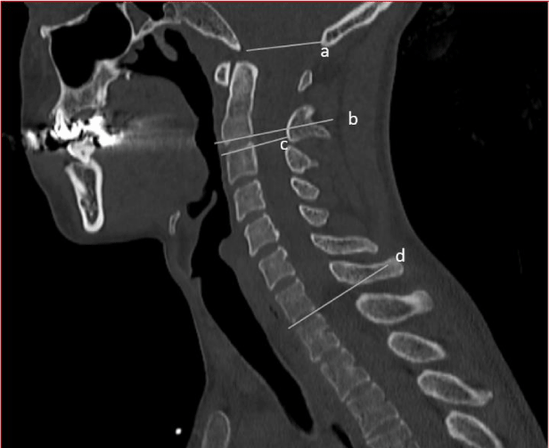
CT evaluations: All evaluations were performed by the same examiner using the multi-planar reconstruction feature to better reposition the slices according to the anatomical plans in addition to obtaining accurate cervical angles. Four lines were drawn on each mid-sagittal CT image (Figures 1,2):
- Foramen magnum (FM-C0): between the basion and the opisthion
- C2: parallel to the inferior endplate of C2
- C3- parallel to the superior endplate of C3
i. Using these four lines the following lordosis measurements were performed in the midsagittal plane (Figure Total cervical lordosis - (foramen magnum C0-C7) Cobb angle of full cervical between the foramen magnum to the C7 inferior endplate.
ii. Upper cervical lordosis- (foramen magnum C0-C3) Cobb angle of the upper cervical between the foramen magnum and the C3 superior endplate.
iii. Mid-lower cervical lordosis - (C2-C7) - Cobb angle between the C2 inferior endplate and the C7 inferior endplate.
iv. Lower cervical lordosis (C3-C7)- Cobb angle between the C3 superior endplate and the C7 inferior endplate.
The above measurements have been commonly used in cervical lordosis studies [7]. By utilizing these four measurements, it became possible to compare the current results with lordosis angles obtained by other researchers.
Reliability of measurements: One assessor who read and re-read 20 CTs, 7 days apart, was blinded to the identity of the patient to assess the intra-rater reliability of the Cobb angle measurements. Intraclass correlations (ICC) were calculated on repeated measurements to assess intratester reliability, with results between 0.87 and 0.91, demonstrating excellent reliability.
Statistical analysis: The differences between groups (individuals with and without a history of cervical trauma) were examined by the Student’s t-test (for continuous variables) and the χ2-test (for categorical variables). The sex differences of cervical angles were examined by independent-sample t-test analysis. One-way ANOVA was used to compare the four cervical lordosis measurements between three age groups by sex. The univariate analysis explored the associations between background and professional characteristics (age, sex, BMI, years working in the profession, working hours, regular physical activity, sedentarity, and light work) and the cervical lordosis measurements. The predictors that showed a significant (p<0.05) association with cervical lordosis measurements were included in the multivariate regression analysis. Linear regression analyses evaluated the association between all background and professional characteristics, and separately for each of the four measurements of cervical angles. A backward procedure was used with the entry probability of 0.05. All independent variables with significant associations were included in the final model. Data were analyzed using SPSS 23.0 for Windows.
Results
Demographic and professional characteristics are shown in Table 1. Individuals with a history of cervical trauma and those without a history of accidents significantly differed in their professional characteristics: age (mean 54.47±12.81 vs 46.90±15.16), sedentarity (20.7% vs 35.0%), and light work (34.7% vs 53.4%). No significant differences were found in BMI, years working in the profession, working hours per week, sex, and regular physical activity (at least two hours a week).
Mean and standard deviations of cervical angles by age (three age groups) and sex are presented in Table 2 (subjects without cervical trauma) and Table 3 (subjects with a history of cervical trauma). In subjects without cervical trauma (Table 2), males showed significantly high (C3-C7) (p= 0.023) cervical lordosis. A comparison between the three age groups (one-w er angles of cervical lordosis compared to females (t-test) aged 20-39, in lower (C2-C7) (p= 0.006) and mid-lower (C3-C7) (p= 0.017) cervical lordosis. Males aged 40-59 showed significantly higher angles of cervical lordosis in the total (C0-C7) (p= 0.021), mid-lower (C2-C7) (p= 0.031), and lower ay ANOVA), revealed a statistically significant increase of cervical lordosis only in females, in the mid-lower (C2-C7) (F2= 5.86; p= 0.005) and lower (C3-C7) (F2= 8.12; p= 0.001) lordosis.
In subjects with a history of cervical trauma (Table 3), males showed significantly higher angles of cervical lordosis compared to females (t-test) aged 20-39, in the mid-lower (C2-C7) lordosis measurement (p= 0.021). In males aged 40-59, significant differences were found in the mid-lower (C2-C7) (p= 0.011) and lower (C3-C7) (p= 0.003) cervical lordosis. A comparison between the three age groups and cervical angles (one-way ANOVA) revealed a statistically significant increase of cervical lordosis in males in the total (C0-C7) (F2= 5.46; p= 0.007), mid-lower (C2-C7) (F2=3.46; p=0.040) and lower (C3-C7) (F2= 4.07; p= 0.023) lordosis. In females, a statistically significant increase of cervical lordosis was found in the total (C0-C7) (F2= 3.87; p= 0.028), mid-lower (C2-C7) (F2= 5.90; p= 0.005) and lower (C3-C7) (F2= 5.49; p= 0.008) lordosis.
In the linear regression analyses (Table 4) with total cervical lordosis (C0-C7) as a dependent variable, the following predictors were found statistically significant: age (β= 0.28, p <0.001), sex (β= 0.23, p= 0.001), and a history of cervical trauma (β= 0.15, p= 0.035). The proportion of the variance explained by the model was 17% (Nagelkerke R-square=0.17). The results of the linear regression analyses for upper cervical lordosis (C0-C3) revealed the following significant predicting variables: sedentary work (β= 0.17, p= 0.019), and BMI (β= 0.14, p= 0.045). The proportion of the variance explained by the model was 7.2% (Nagelkerke R-square =0.072). The results of the linear regression analyses for mid-lower cervical lordosis (C2-C7) revealed the following significant predicting variables: age (β= 0.25, p<0.001), sex (β= 0.33, p<0.001), history of cervical t rauma (β= 0.16, p= 0.019), and sedentary work (β=-0.18, p= 0.008). The proportion of the variance explained by the model was 21% (Nagelkerke R-square=0.21). The results of the linear regression analyses for lower cervical lordosis (C3-C7) revealed the following significant predicting variables: age (β= 0.25, p<0.001), sex (β= 0.27, p<0.001), and sedentary work (β= -0.16, p= 0.023). The proportion of the variance explained by the model was 16% (Nagelkerke R-square= 0.16).
Discussion
Association of cervical lordosis with a history of cervical trauma
Herein, it was found that a history of cervical trauma was associated with total cervical (C0-C7) and mid-lower lordosis (C2-C7), indicative of a lordosis reduction. The current results concur with Marshall & Tuchin’s conclusions [19], of a retrospective analysis of 500 radiographs examining the correlation between cervical lordosis (C1-C7 angle) and history of a motor vehicle accident. They found that cervical lordosis was reduced in most (82%) of the patients with a history of a motor vehicle accident. Beltsios, et al. [5], note that accidents can damage the cervical spine, and lead to degenerative changes, i.e., decreased intervertebral disc height and muscle weakness. Gao, et al. [20], evaluated the correlation between cervical lordosis and cervical disc herniation in 300 patients with neck pain, under the age of 40. For this purpose, X-rays were taken with the patients in a standing position. They found that the degree of herniation was higher in the straight and kyphosis groups compared to the lordosis group. In the current study, similarly, cervical trauma was associated with decreasing in cervical lordosis.It is concluded that a history of cervical trauma is an important reason for the occurrence of degenerative changes leading to changes in cervical lordosis, probably decreasing lordosis, and should be examined when researching cervical spine morphology and degeneration.
Association of cervical lordosis with age and sex
The current study reveals that age and sex are associated with mid-lower (C2-C7) and lower (C3-C7) cervical lordosis. No association was found with the upper cervical lordosis angles. Furthermore, significant sex differences were found only in the young (20-39) and intermediate (40-59) age groups, with lordotic angles greater in men. When comparing the three age groups (20-39, 40-59, 60+), differences were found in the groups without cervical trauma in the mid-lower and lower cervical lordosis. Only in females, increased cervical lordosis with age was observed.
There have been contrasting reports as to the association between cervical alignment and age in the elderly. It has been reported that a noticeable increase in cervical lordosis occurs with age [21]. A recent study reported that owing to aging, the mid-lower cervical lordosis (C2-C7) increases from 8° (30 years old) to 20° (80 years old). Other reports have confirmed a lordosis angle decrease as people age, with the development of a kyphotic cervical curve [22]. An additional study [7] has found a similar total cervical lordosis (C0-C7) in both adult males and females (20-50 years of age), however, several significant differences between sexes were discovered. Females exhibited a higher (by 5°) upper cervical lordosis (C0-C3; C1-C3) than males, whilst, males displayed a higher lower cervical lordosis (C3-C7) than females (by 6°). These conclusions concur with Yukawa, et al. [21] findings that a greater mid-lower cervical lordosis (C2-C7) exists in males compared with females aged 30-80.
Several studies reported differences found in the cervical lordosis angle due to age and sex [23]. Yukawa, et al.’s [21] conclusions were similar to ours, i.e., female cervical lordosis increases with age, however, the angle is greater in males. X-ray imaging was used while standing. The subjects were all Japanese. Cervical lordosis was measured only at the C2-C7.
Relevant studies (16,21,26,30,31), comparing their C2-C7 cervical lordosis Cobb angle values (8.21o±14.00o) in symptomatic individuals with the current study, are summarized in Table 5. Our measurements of C2-C7 are the lowest, similar to McAviney, et al. [24] results, although the latter used x-rays as a measurement. Our results, in accordance with Jun, et al.’s [17], demonstrated lower cervical lordosis values while lying supine (by CT scans) compared with imaging in a standing position (by x-rays) of the C2-C7. Only two modalities were used [17], CT scans, and x-rays. The authors discovered that cervical lordosis Cobb angles C2- C7 were significantly smaller than when measured by x-rays. The authors included only 50 individuals of all ages.
Association of cervical lordosis with occupational variables
In the current study, it was found that sedentarity was positively associated with lordosis of the upper cervical spine and negatively with the lower. This interesting outcome, as yet unpublished elsewhere, may be related to sedentarity and neck pain, a subject discussed in the literature. In a review was reported that sitting for long periods and holding the head in a forward position, causes cervical sagittal imbalance, and furthers the development of cervical pain and pathology [6]. Ames, et al. [25], suggest that cervical posture affects the development of cervical pathology, however, if this relationship exists, it has not yet been fully acknowledged.
Workers in various professions such as office personnel [15,16], ultrasonographers [26], dental hygienists [27], and professional drivers [13,14] mainly suffer from neck pain. Forward head posture, weekly computer use for ≥6-9 hours, continuous sitting, incorrect placement of computer devices, i.e., the monitor, keyboard and mouse, and the position of the keyboard, have been associated with the prevalence of neck pain [15,16]. Furthermore, an uncomfortable steering wheel and/or seat and back support were found associated with a higher prevalence of neck pain in professional urban bus drivers [13]. Driving with a bent or twisted trunk was associated with neck pain [14]. Sonographers, whose screen is on their left side, experience significantly more neck pain [28].
The strongest risk factor for neck pain in females is sustained/repeated arm abduction with high physical exertion. A higher incidence of neck pain in men was due to protracted forward head flexion [29].
Two studies have shown that degenerative changes, i.e., decreased vertebral body, intervertebral disc height, laxity and muscle weakness, together with neck flexion and neck loads over time, can lead to changes in cervical lordosis [3,5]. A forward head posture develops when working in a sedentary position [30], which may lead to neck and head pain [16]. This type of sedentary work including static neck bending mechanisms will likely lead to decreased upper and increased lower cervical lordosis. It has been recently reported that there are ergonomic solutions for maintaining good body posture, thus, preventing neck loads with pain [31-33]. These prevention strategies should be available to sedentary workers.
Limitations
Our study has several limitations: (1) our subjects were scanned in a supine position implying a different gravity effect on the neck, compared with standing position. Although other similar studies positioned their patients in a standing or a sitting position, recent studies have used the supine position for the evaluation of cervical lordosis [17,18]. Another important reason for using the supine position is the increasing use of CT and MRI scans for evaluation of neck pain and cervical pathology [17,18]; (2) our study included a convenience sample rather than a representative one. Moreover, all subjects experienced neck-related symptoms. Healthy subjects may show different results; (3) no data were available as to the time since the accident, the nature of the accident and its severity for the research group with a history of cervical trauma.
Conclusion
There is a difference in cervical lordosis between individuals suffering from neck pain, with and without a history of cervical trauma. It is most likely that degenerative changes in the cervical spine following cervical accidents, lead to changes in the cervical lordosis. The minimum potential period between the accident and the presence of these changes is unknown and should be investigated as well. Mid-lower and lower cervical lordosis in young and middle-aged individuals are higher in males compared with females, but at an older age, no differences were found between the sexes. Future research is needed to assess the association between age-related change in sex hormone levels and changes in cervical lordosis. Do women have a greater kyphosis in old age, thereby compensating for increased cervical lordosis? In the present study, a correlation was found between sedentarity, and the upper, mid-lower, and lower cervical lordosis, a new finding. Intervention studies aimed at improving posture to prevent degenerative changes and pain in sedentary workers should be continued. Prevention strategies should be available to sedentary workers to maintain proper lower neck lordosis and prevention of upper neck lordosis exaggeration.
Author contribution
D EZRA: Project development, Data Collection, Data analysis, Manuscript writing
L KALICHMAN: Project development, Data analysis, Manuscript writing
A SIMONOVICH: Project development, Data Collection
J DROUJIN: Project development, Data Collection
E BEEN: Project development, Manuscript writing
D ALPEROVITCH-NAJENSON: Project development, Data Collection, Data analysis, Manuscript writing
Conflict of interest statement
All authors participated in the design and conduct of the study, have read and approved the manuscript, meet the requirements of authorship, have no conflict of interest, consider the manuscript to present honest work and have respected all ethical principles of the World Medical Association and the Declaration of Helsinki.
The authors would like to thank the participants referred for a CT to Barzilai Medical Center, Ashkelon, Israel, for their cooperation in data collection, and Mrs Phyllis Curchack Kornspan for her editorial services.
- Yoon SY, Moon HI, Lee SC, Eun NL, Kim YW (2018) Association between cervical lordotic curvature and cervical muscle cross‐sectional area in patients with loss of cervical lordosis. Clin Anat 31: 710-715. Link: https://bit.ly/2Y6Mcti
- Miyazaki M, Hong SW, Yoon SH, Morishita Y, Wang JC (2008) Reliability of a magnetic resonance imaging-based grading system for cervical intervertebral disc degeneration. J Spinal Disord Tech 21: 288-292. Link: https://bit.ly/2yPfh1z
- Ezra, D, Masharawi Y, Salame K, Slon V, Alperovitch-Najenson D, et al. (2017) Demographic aspects in cervical vertebral bodies' size and shape (C3–C7): a skeletal study. Spine J 17: 135-142. Link: https://bit.ly/3aHblNQ
- Scheer JK, Tang JA, Smith JS, Acosta FL, Protopsaltis TS, et al. (2013) Cervical spine alignment, sagittal deformity, and clinical implications: a review. J Neurosurg Spine 19: 141-159. Link: https://bit.ly/2VFGMUr
- Beltsios M, Savvidou O, Mitsiokapa EA, Mavrogenis AF, Kaspiris A, et al. (2013) Sagittal alignment of the cervical spine after neck injury. Eur Orthop Surg Traumatol 23: S47-S51. Link: https://bit.ly/2SdnvaV
- Ezra, D, Been, E, Alperovitch-Najenson, D, Kalichman L (2019 ) Cervical posture, pain, and pathology: developmental, evolutionary and occupational perspective. In Spinal Evolution Springer, Cham 321-339. Link: https://bit.ly/3cLDDbe
- Been E, Shefi S, Soudack M (2017) Cervical lordosis: the effect of age and gender. Spine J 17: 880-888. Link: https://bit.ly/3aKGe3L
- Ariëns GA, Bongers PM, Douwes M, Miedema MC, Hoogendoorn WE, et al. (2001) Are neck flexion, neck rotation, and sitting at work risk factors for neck pain? Results of a prospective cohort study. Occup Environ Med 58: 200-207. Link: https://bit.ly/2VZFLpk
- Côté P, van der Velde G, Cassidy JD, Carroll LJ, Hogg-Johnson S, et al. (2008) The burden and determinants of neck pain in workers: results of the Bone and Joint Decade 2000-2010 Task Force on Neck Pain and Its Associated Disorders. Spine 15: 33: S60-S74. Link: https://bit.ly/3eVMB7W
- Mousavi-Khatir R, Talebian S, Toosizadeh N, Olyaei GR, Maroufi N (2018) The effect of static neck flexion on mechanical and neuromuscular behaviors of the cervical spine. J Biomech 72: 152-158. Link: https://bit.ly/3aG8zs3
- Ranasinghe P, Perera YS, Lamabadusuriya DA, Kulatunga S, Jayawardana N, et al. (2011) Work related complaints of neck, shoulder and arm among computer office workers: a cross-sectional evaluation of prevalence and risk factors in a developing country. Environ Health 10: 70. Link: https://bit.ly/2KBS8Cv
- Hush JM, Michaleff Z, Maher CG, Refshauge K (2009) Individual, physical and psychological risk factors for neck pain in Australian office workers: a 1-year longitudinal study. Eur Spine J 18: 1532-1540. Link: https://bit.ly/2W5L2LQ
- Alperovitch-Najenson D, Katz-Leurer M, Santo Y, Golman D, Kalichman L (2010) Upper body quadrant pain in bus drivers. Arch environ occup health 65: 218-223. Link: https://bit.ly/2yQtXxq
- Bovenzi M (2015) A prospective cohort study of neck and shoulder pain in professional drivers. Ergonomics 58: 1103-1116. Link: https://bit.ly/2Y9Xptc
- Jun D, Zoe M, Johnston V, O’Leary S (2017) Physical risk factors for developing non-specific neck pain in office workers: a systematic review and meta-analysis. Int Arch of Occup Environ Health 90: 373-410. Link: https://bit.ly/2W0a5QC
- Nejati P, Lotfian S, Moezy A, Nejati M (2015) The study of correlation between forward head posture and neck pain in Iranian office workers. Int J Occup Med Environ Health 28: 295-303. Link: https://bit.ly/2KBeyUx
- Jun HS, Chang IB, Song JH, Kim TH, Park MS, et al. (2014) Is it possible to evaluate the parameters of cervical sagittal alignment on cervical compute tomographic scans? Spine 39: E630-E636. Link: https://bit.ly/2Sbylhx
- Liu W, Fan J, Bai J, Tang P, Chen J, et al. (2017) Magnetic resonance imaging: A possible alternative to a standing lateral radiograph for evaluating cervical sagittal alignment in patients with cervical disc herniation? Medicine 96: e8194. Link: https://bit.ly/3bFZ7pT
- Marshall DL, Tuchin PJ (1996) Correlation of cervical lordosis measurement with incidence of motor vehicle accidents. Australas Chiropr Osteopathy 5: 79-85. Link: https://bit.ly/2VYy2rq
- Gao K, Zhang J, Lai J, Liu W, Lyu H, Wu Y, Lin Z, Cao Y (2019) Correlation between cervical lordosis and cervical disc herniation in young patients with neck pain. Medicine (Baltimore). 98: e16545. Link: https://bit.ly/2Y7uFB7
- Yukawa Y, Kato F, Suda K, Yamagata M, Ueta T (2012) Age-related changes in osseous anatomy, alignment, and range of motion of the cervical spine. Part I: Radiographic data from over 1,200 asymptomatic subjects. Eur Spine J 21: 1492-1498. Link: https://bit.ly/35ayDe0
- Boyle JJ, Milne N, Singer KP (2002) Influence of age on cervicothoracic spinal curvature: an ex vivo radiographic survey. Clin Biomech 17: 361-367. Link: https://bit.ly/3aH7fVK
- Kasai T, Ikata T, Katoh S, Miyake R, Tsubo M (1996) Growth of the cervical spine with special reference to its lordosis and mobility. Spine 21: 2067-2073. Link: https://bit.ly/3cNlGcz
- McAviney J, Schulz D, Bock R, Harrison DE, Holland B (2005) Determining a clinical normal value for cervical lordosis. J Manipulative Physiol Ther 28: 187-193. Link: https://bit.ly/3eSrh32
- Ames CP, Blondel B, Scheer JK, Schwab FJ, Le Huec JC, et al. (2013) Cervical radiographical alignment: comprehensive assessment techniques and potential importance in cervical myelopathy. Spine 38: S149-S160. Link: https://bit.ly/3593ozL
- Claes F, Berger, J, Stassijns G (2015) Arm and neck pain in ultrasonographers. Human factors 57: 238-245. Link: https://bit.ly/2zz4HfM
- Pettit NJ, Auvenshine RC (2018) Change of hyoid bone position in patients treated for and resolved of myofascial pain. Cranio 38: 74-90. Link: https://bit.ly/2VWV3Lo
- Hayes MJ, Osmotherly PG, Taylor JA, Smith DR, Ho A (2016) The effect of loupes on neck pain and disability among dental hygienists. Work 53: 755-762. Link: https://bit.ly/2VGZafu
- Kang JH, Park RY, Lee SJ, Kim JY, Yoon SR, et al. (2012) The effect of the forward head posture in long time computer-based workers. Ann Rehabil Med 36: 98-104. Link: https://bit.ly/357KmKd
- Ailneni RC, Syamala KR, Kim IS, Hwang J (2019) Influence of the wearable posture corecction sensor on head and neck posture: Sitting and standing work stading workstations. Work 62: 27-35. Link: https://bit.ly/2KA3w1F
- Núñez-Pereira S, Hitzl W, Bullmann V, Meier O, Koller H (2015) Sagittal balance of the cervical spine: an analysis of occipitocervical and spinopelvic interdependence, with C-7 slope as a marker of cervical and spinopelvic alignment. J Neurosurg Spine 23: 16-23. Link: https://bit.ly/3aBpTyl
- Shilton M, Branney J, de Vries BP, Breen AC (2015) Does cervical lordosis change after spinal manipulation for non-specific neck pain? A prospective cohort study. Chiro Man Therap 23: 33. Link: https://bit.ly/35aygA8
- Tang JA, Scheer, JK, Smith JS, Deviren V, Bess S, et al. (2012) The impact of standing regional cervical sagittal alignment on outcomes in posterior cervical fsion surgery. Neurosurgery 71: 662-669. Link: https://bit.ly/2y08hPq
Article Alerts
Subscribe to our articles alerts and stay tuned.
 This work is licensed under a Creative Commons Attribution 4.0 International License.


 Save to Mendeley
