Journal of Novel Physiotherapy and Physical Rehabilitation
Optimizing Rehabilitation: The Potential to Assess Cardiorespiratory, Neuromuscular and Biomechanical Adaptations to Exercise of Children with Cerebral Palsy in the Face of Intra-Individual Variation
Angeline N Leunkeu1*, Roy J Shephard2 and Said Ahmaidi1
2Faculty of Kinesiology and Physical Education, University of Toronto, PO Box 521, Brackendale, BC V0N 1H0, Canada
Cite this as
Leunkeu AN, Shephard RJ, Ahmaidi S (2017) Optimizing Rehabilitation: The Potential to Assess Cardiorespiratory, Neuromuscular and Biomechanical Adaptations to Exercise of Children with Cerebral Palsy in the Face of Intra-Individual Variation. J Nov Physiother Phys Rehabil 4(2): 042-047. DOI: 10.17352/2455-5487.000045The aim of this article is to assess the practical clinical value of measures used to examine physiological (cardiorespiratory and neuromuscular) and biomechanical (spatio-temporal and baropodometric) responses to effort in children with cerebral palsy (CP). Cardiorespiratory data included a 6 minute walking test and cycle ergometric determinations of peak respiratory gas exchange V( O2peak,VE,) by Cosmed K4b2 gas analyzer. Peak isometric strength and fatigue for the quadriceps muscle were tested on the Cybex (Norm II) isokinetic apparatus, noting the time required for effort to decrease to 50% of the maximal voluntary isometric force (MVIF). Qualitative analysis of the associated electromyographic signal examined the root mean square voltage and median frequency. Biomechanical data included gait cycle characteristics (speed, step length, step frequency, impulse, time of contact, step duration, time of double support) and plantar pressure peaks measured at a total of 8 sites. Values for all of these variables met or exceeded conventional standards of reliability and validity, but a substantial intra-individual test-retest variation limited the potential to interpret changes in an individual’s physical condition. The authors emphasize that whether looking at aerobic function, muscular fatigue or gait, it is necessary to gauge the effectiveness of training programmes in terms of grouped rather than individual responses.
Introduction
An optimal rehabilitation programme is based upon a clear diagnosis of functional deficits, an understanding of the potential for reversing these deficits, and effective procedures to monitor the intensity of rehabilitation and determine the progression of clinical condition. Critical to this process are the reliability and validity of the physiological measuring tools that are chosen relative to the likely gains of condition that an individual will make during rehabilitation. In children with cerebral palsy, a varying amount of muscle spasm limits is particularly liable to reduce both the repeatability of test results and likely gains in aerobic power, muscular strength and gait with training. This article thus examines critically practical methods for evaluating cardio-respiratory fitness, strength and gait in children with cerebral palsy. Specifically, it looks at the reliability and validity of individual techniques, and considers their ability to detect clinically meaningful changes of function in children with cerebral palsy. We first describe the test sample used in our investigation and general issues of methodology, and then report findings for each of these 3 areas of assessment.
General methodology
Study design: A group of 14-year-old children with cerebral palsy (Ashworth gross motor classification system, levels I and II, Table1) were first familiarized with the test measurements of interest. For the aerobic tests, definitive data were obtained before and after an 8-week period in which a half of the 28 participants were assigned to a training programme (40 minutes of moderate walking exercise, 3 times/week), and a half served as a matched control group. Strength and gait measurements were obtained on similarly sized groups of children with hemiplegic and bilateral cerebral palsy (n = 12), with repeat assessments after an interval of 1 week.
Participants: A total of 28 children were initially recruited in accordance with a protocol approved by the Human Experimentation Committee of the University of Amiens. Participants and their parents gave formal consent to the procedures, on the understanding that withdrawal from the study would not affect their subsequent treatment. Four of 28 children did not complete the investigations. Two of these were obliged to stop training because of pending surgery, and the other 2 children were unwilling to wear a face-mask. Thus, 24 participants completed the entire training study.
Tests of aerobic function: The aerobic tests included a 6-min walking test (6mwt) and a ramp cycle ergometer stress test (increments of 15 W per min) performed to voluntary exhaustion on a Monark 824 ergometer (Monark, Vansbro, Sweden). Direct measurements of peak oxygen intake were made using a Cosmed K4b2 portable gas analyzer (Cosmed, Rome, Italy).
In the 6mwt, participants covered the maximum possible distance in 6 minutes, walking along a hospital corridor. We recorded the distance walked, the peak heart rate, the peak ventilation, and an estimate of peak oxygen intake (the highest value observed during the final 5 seconds of testing).
Tests of muscular strength and fatigue: Muscle strength and fatigue were assessed on a Cybex isokinetic dynamometer at knee angles approximating 60° and 120° (depending on the individual’s limb flexibility). We made 5 determinations of maximal voluntary force, and timed the ability to hold contractions within 5% of 50% of maximum force. Surface electromyographic measurements were made over the active muscles.
Tests of gait: Gait was assessed using 24 Parotec in-shoe pressure sensors (Paromed Medizintechnik GmbH., Neubeuern, Germany). Data reproducibility was assessed by two walking trials (4 X 12 meters at self-selected speeds) performed with a one-week inter-test interval. Spatio-temporal gait cycle parameters included step duration (Tstep), double-support time (Tds), ground contact time (Tc), walking velocity (V), step frequency (F), and stride length (A). Peak plantar pressures were recorded at 8 sites on the soles of the feet: lateral heel, medial heel, lateral mid-foot, medial mid-foot, fourth and fifth metatarsal heads, second and third metatarsal heads, first metatarsal head, and hallux.
Statistical analysis
Conventional measures of test reliability and validity were determined using StatView Software (SAS Institute Inc, SAS Campus Dr, Cary, North Carolina 27513, USA). Statistical significance was set at p<0.05 throughout. Individual data were expressed as mean values ± standard deviation (SD). Comparisons were made by analysis of variance (ANOVA), and a Schéffé post-hoc test located significant differences. Test-retest reliability, using the same examiner, was determined using Spearman correlation coefficients (r), intraclass correlation coefficients (ICCs) and Bland-Altman plots [1]. 95% of the data for both the aerobic endurance tests and isometric strength parameters fell within the limits of the corresponding Bland-Altman plots, as required in data of acceptable repeatability [1]. Assuming the mean difference between the first and second measurements to be zero, the COR for the Bland-Altman test was derived as follows.
Where x1 and x2 are the mean values for the first and second measurements and n is the number of subjects. Bland-Altman plots were generated, demonstrating satisfactory relationships between inter-measurement differences and their average, together with the limits of agreement (mean difference, ±2SDs) [1,2].
Results
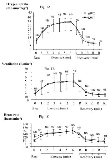
Aerobic function: Data obtained for the 6 mwt showed good reliability (Figure 1); for V O2peak: test/retest correlation r=0. 90, p<0.001, ICC=0.85; for peak ventilation: r=0.88, p<0.001, ICC=0.83; for peak heart rate: r=0.86, p<0.001, ICC=0.82; and for distance walked: r=0.87; p=0.007, ICC=0.80.
The validity of the test as an index of aerobic function was shown by similar mean values and a fairly close correlation of individual mwt data with the corresponding cycle-ergometer values (6mwt vs. cycle ergometer peak oxygen intake, r = 0.63). Significant improvements in 6mwt V O2peak, peak ventilation and peak heart rate were found after 8 weeks of training (p<0.05), with no changes in the performance of control participants.
Muscular strength and fatigue
The Cybex data showed satisfactory reliability, with very similar values for maximum voluntary strength and endurance at 50% of maximum strength before and after the 1-week interval (r=0.87, p<0.01, ICC=0.84). The data were adequate to demonstrate that children with right-sided hemiplegic CP developed a significantly weaker maximum quadriceps force in their affected leg than in their control leg (p < 0.0001) and in their unaffected leg versus the force developed by the corresponding leg in control participants (p<0.01). However, perhaps because the children with hemiplegic CP made a less complete maximal effort, and/or that the affected limb was fatiguing from a lower level of activity, no significant differences in contraction duration were found between the affected and unaffected legs (p=0.26), or between the unaffected leg and that of control participants (p= 0.67).
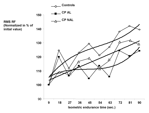
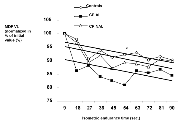
In support of the idea that children with hemiplegic CP made a lesser maximal effort, the normalized root mean square emg voltages over the rectus femoris were significantly lower for the affected leg (AL) than for the control leg (CL) (Figure 2). The slope of the decline in force [as calculated by Portero et al. [3] for the vastus lateralis] was also greater for the affected than for the unaffected leg (Figure 3).
Gait and plantar pressures
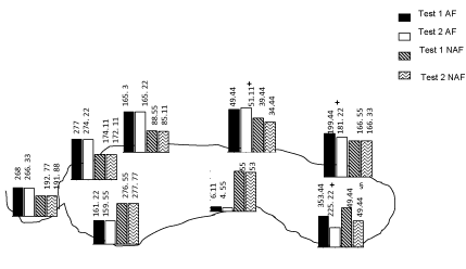
Spatio-temporal parameters for the unaffected limb showed very similar mean values for tests 1 and 2, with no significant inter-test differences (Figure 4). Correlations between paired data sets were statistically significant, except for contact time [V: r = 0.75, P = 0.009; F: r = 0.95, p<0.0001; A: r = 0.59, p = 0.05; Tds: r = 0.94, p <0.0001; Tstep: r = 0.93, p <0.0001) and Tc: r = 0.41, p = 0.13]. Given the reliability of gait measurements, significant differences of walking speeds (m∙s-1) were shown between subject groups (0.65 ± 0.13 for hemiplegia, 0.93 ± 0.22 for diplegia and 1.26 ± 0.05 for able-bodied children), stride lengths being shorter in those with cerebral palsy. Contact time, double support time and step duration were also significantly shorter in those individuals with hemiplegia.
Peak pressures for the unaffected limb showed no statistically significant inter-test differences, except under the medial heel (p<0.0001), where values were much lower at the second assessment. All repeat measurements reached an ICC > 0.84, with data for lateral mid-foot, fourth and fifth metatarsal heads and hallux in the range 0.97-0.99 (Figure 4). Moreover, 95% of the data for most of the plantar pressures fell within the limits set by the corresponding Bland-Altman plots (Figure 5). Spearman and intra-class correlations were also high for several measurements on the affected limbs: (lateral heel: 0.99 and 0.99; medial heel: 0.97 and 0.96; lateral mid-foot: 0.90 and 0.90; fourth and fifth metatarsal heads: 0.95 and 0.95; and hallux: 0.87 and 0.77), although peak pressures showed significant inter-test differences in most foot regions (p<0.05).
Plantar pressures differed substantially and consistently between able-bodied children and those with cerebral palsy, with increased medial heel pressures in hemiplegia, and reduced hallux and lateral heel pressures but increased lateral, medial mid-foot and first metatarsal pressures in children with diplegia.
Discussion
This article draws summarizes our practical experience in using laboratory tests to optimize the cardio-respiratory function, muscular strength and gait of children with cerebral palsy. In particular, it looks critically at the reliability and validity of current methodology [2,4,5]. Relative to the magnitude of changes induced by the disease process and by participation in a modest exercise-centred rehabilitation programme.
At first inspection, all of the laboratory measurements might be judged as very satisfactory, since they meet traditionally accepted criteria of reliability and validity. Thus, the 6mwt provides a group estimate of O2peak that is comparable to laboratory-base cardiopulmonary ergometer data in children with spastic cerebral palsy (GMFCS levels I and II) [7]. The cardiorespiratory responses seen during the two forms of testing are also similar and correlate closely with each other. Moreover, the 6WT provides closely repeatable data over a one-week interval, and has construct validity in terms of demonstrating group adaptations of cardiorespiratory function and physical performance over 8 weeks of moderate intensity training [5,6,8]. We may conclude that the 6mwt is a reproducible procedure, with a validity demonstrated by a correlation with data obtained by cycle-ergometer assessments of cardio-respiratory response. It thus offers a simple basis for the assessment of changes in the aerobic function of small groups of children with cerebral palsy. But although the group response demonstrates the efficacy of rehabilitation, as with most clinical tests, the test-retest variance is too large to form a reliable impression of an individual’s training response relative to the likely gains in aerobic function.
Likewise, the measurements of isometric strength show good reproducibility, with a test-retest reliability that is sufficient to demonstrate significant inter-group differences in performance, but not enough to define clearly changes of strength in any given child. Moreover, it is somewhat surprising that the time to fatigue did not differ for the diseased limbs. One possible explanation of this finding could be that the children with CP did not accomplish a true maximual effort, whether because of weaker motivation, or because of co-contraction of antagonist muscles [9]. In support of this view, Rose et al. [10]. determined that neuromuscular activation during maximum voluntary contraction was significantly reduced in subjects with CP when compared with controls, and that individuals with CP were unable to recruit higher threshold motor units or to drive lower threshold motor units to a higher firing rate. If the subjects with CP were not able to attain a true maximual effort, then they were not working at an equal percentage of the MVIF compared to the able-bodied individuals.
Despite the value of these various tests in demonstrating both group differences associated with the underlying disease and responses to exercise rehabilitation, it must be underlined that with all of these measures, there were substantial intra-individual test-retest differences that limited their value in advising the individual patient. Although not widely acknowledged, this is an important limitation to almost all of the procedures used in clinical physiology at the present time. When using laboratory tests, the focus should thus be on obtaining group data that can be used to assess the efficacy of procedures, rather than engage in an over-interpretation of apparent changes in an individual’s test scores.
Conclusions
Our research has demonstrated the effectiveness of a battery of clinical tests to provide clinically useful information on the group efficacy of programmes for the rehabilitation of children with cerebral palsy. Moderate walking, practised three times a week for 8 weeks, is able to produce beneficial outcomes for children with CP in terms of enhanced gas exchange, gains in strength and improved walking performance. Despite the valuable information that can be obtained concerning group responses, care must be taken in interpreting apparent changes within an individual patient, since the intra-individual test-retest variance is substantial relative to the likely effects of either the disease process or the response to rehabilitation.
- Bland JM, Altman DG (1986) Statistical methods for assessing agreement between two methods of clinical measurement. Lancet 8: 307-310. Link: https://goo.gl/Nxbx1e
- Bland JM, Altman DG (1990) A note on the use of interclass correlation coefficient in the evaluation of agreement between two methods of measuremnts. Comput Biol Med 20: 337-340. Link: https://goo.gl/FA2mRD
- Portero P, Vanhoutte C, Goubel F (1996) Surface electromyogram power spectrum changes in human leg muscles following 4 weeks of simulated microgravity. Eur J Appl Physiol 73: 40-45. Link: https://goo.gl/jYckbs
- Nsenga LA, Lelard T, Shephard RJ, Doutrellot PL, Ahmaidi S (2014) Reproducibility of gait cycle and plantar pressure distribution assessment in children with cerebral palsy. Neurorehabilitation 35: 597-606. Link: https://goo.gl/nwUewh
- Nsenga LA, Shephard RJ, Ahamaidi S (2012) Six-Minute Test in children with cerebral palsy Gross Motor Function Classification System Levels I and II: reproducibility, validity, and training effects. Arch Phys Med Rehabil 93: 2333- 2339. Link: https://goo.gl/ySI4mX
- Nsenga LA, Lelard T, Shephard RJ, Doutrellot PL, Ahmaidi S (2014) Gait cycle and plantar pressure distribution in children with cerebral palsy: Clinically useful outcome measures for management and rehabilitation. Neurorehabilitation 35: 657-663. Link: https://goo.gl/N3tISB
- Palisano RJ, Hanna SE, Rosenbaum PL, Russell DJ, Walter SD (2000) Validation of a model of gross motor function for children with cerebral palsy. Phys Ther 80: 974-985. Link: https://goo.gl/FBH5w1
- Nsenga LA, Shephard RJ, Ahmaidi S (2012) Aerobic Training in Children with Cerebral Palsy. Int J Sports Med 33: 1-5. Link: https://goo.gl/BkX1LK
- Nsenga LA, Keefer JD, Miladi I, Ahmaidi S (2010) Electromyographic (EMG) Analysis of quadriceps femoris muscle fatigue in children with cerebral palsy during a sustained isometric contraction. J Child Neurol 25: 287-293. Link: https://goo.gl/GXLV0B
- Rose J, McGill KC (2005) neuromuscular activation and motor-unit firing characteristics in cerebral palsy. Dev Med Child Neurol 47: 329-336. Link: https://goo.gl/9V0mLL
- Ashworth B (1964) Preliminary trial of carisoprodol in multiple sclerosis. Practitioner 192: 540-542 . Link: https://goo.gl/ZYCBJr
Article Alerts
Subscribe to our articles alerts and stay tuned.
 This work is licensed under a Creative Commons Attribution 4.0 International License.
 Save to Mendeley
