Annals of Molecular and Genetic Medicine
Tumor-stroma cross talk and platelets: Curse of cancers
Ashok Vikey*
Cite this as
Ashok Vikey (2020) Tumor-stroma cross talk and platelets: Curse of cancers. Ann Mol Genet Med 4(1): 018-019. DOI: 10.17352/amgm.000008Introduction
Platelets are essential part of our vascular system, named as thrombocytes and their main role is to stop bleeding; by clumping and aggregating at damaged vascular location. Embryonically the platelets originate in bone marrow from megakaryocytes. These are smallest structures among blood cells, with average dimensions less than 4 microns in size, biconvex or straight ends and normally are colorless without nucleus [1].
Routinely these blood components help in clot formation and repair of tissue after trauma or injury, but on the other hand their close association is noticed in various malignancies and infections. This is possible due to following qualities such as;
a) Attachment on surface: Formation of meshwork at suitable environment, that is helpful during anchorage for travelling cells.
b) Aggregation: This quality helps them to group together during activated form to form meshwork.
c) Agglutination: It is the clumping together of platelets.
The above properties play significant role during distant journey of malignant cells, helping them to withstand mechanical damage [2]. Concept of thromboembolic events and coagulation cascade was highlighted by Armand Trousseau in1865. Recently this is understood that, platelet driven growth factors help growth and proliferation of cancers [3].
Historical background
Around two centuries back platelets became familiar to scientists, after discovery of compound microscope in 1830; by Joseph Jackson Lister. The first picture was made by George Gulliver in 1841, and a year later in 1842; William Addison drew picture of platelet- fibrin clot. However maximum awareness was generated when Lionel Beale first published his sketches of platelets in 1864. In 1865 Max Schultzer identified very small structures, which were smaller than red blood cells and termed as, “spherules”. But actual scientific explanation of platelets was put forward by Dr Richard Hill Norris in 1880.
Previous studies are helpful to understand patho-physiologic association of blood platelets during routine reparative phenomenon as well as carcino- angiogenic concepts respectively [4-6].
Discussion
The aggregating nature of this micro structure of blood plays important role to attract disseminated membrane bound vesicles, later on these Platelet-related microparticles (PMP) alarm to initiate tissue proteins such as cytokine molecules and interleukins. Later on these charged energy driven substances help in growth, proliferation and distant spread of many cancerous cells. Apart from this; platelets also play significant role during venous thromboembolic events and vascular anastomosis [7].
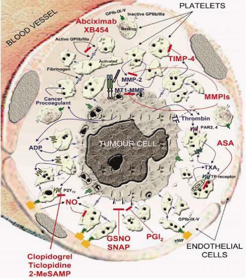
Goubran H. A. et al suggested that, there is direct communication between blood platelets and Extracellular Matrix (ECM) of tumors, as this intra cellular cross-talk activates to release multiple platelet derived growth factors. These charged cellular products orchestrate entire scenario of neoangiogenesis, proliferation and distant spread of malignant cells. Henceforth modification in this molecular concept of platelets and their antagonist companions may be helpful step towards restricting the growing cancers [8].
During process of tumor-stroma cross talk; cancer cells hijack genetic phenotype and remodel growth factors of platelets, which further act as immune mediators during rapid progress of neoplastic cells [9]. Along with this, Cancer cells have skill to change genetic phenotype of extracellular matrix and by this virtue they collect platelets in early stages of metastasis. This collection of platelets is known as Tumor Cell-induced Platelet Aggregation (TCIPA). This is well organized arrangement, made up of many supportive substances such as adrenaline, serotonin and thrombin; those further mediate to thromboxane A2 (TXA2), an important component activating platelet related component GPIIb/IIIa. Later on GPIb-IX-V and GPIIb/IIIa activate platelets to present P-selectins; which are responsible for interaction of leukocytes and platelets. In this manner platelets make cyto-cellular structure to provide safe and comfortable environment to cancer cells for further growth, progress and proliferation (Figure 1) [10].
Conclusion
Platelets are very small but highly significant blood components, as they play crucial role in repair and regeneration during routine physiology. However; on other hand they act as antagonist, in some pathological states such as carcinogenesis, infections and degenerative states of body. Most of cancer related deaths are due to metastasis of cancers, and this is quite possible because of inadvertent nature of platelets and associated growth factors. So understanding this basic patho-physiology of these blood cells, can be guiding force in the field of molecular biology during interventions of various cancers.
Disclosure of potential conflicts of interest: No specific funding was received for the publication of this review. The authors have no conflicts of interest, commercial or otherwise, to declare.
- Sembulingam K, Sembulingam P (2005) Essentials of Medical physiology. 3rd ed. New Delhi: Jaypee brothers medical publishers Ltd.
- Fujita N, Takagi S (2012) The impact of Aggrus / podoplanin on platelet aggregation and tumour metastasis. J Biochem 152: 407-413. Link: https://bit.ly/3ixbWFF
- Goubran HA, Burnouf T, Radosevic M, El-Ekiaby M (2013) The platelet-cancer loop. Eur J Intern Med 24: 393-400. Link: https://bit.ly/2GtAAKv
- Gulliver G (1882) Gulliver’s Researches in Anatomy, physiology, pathology and Botany. Carpenter’s Physiology,ed. Power ed. 9th. Lancet 2: 916.
- Beale LS (1864) On the germinal Matter of the Blood, with Remarks upon the formation of Fibrin”. Transactions of the Microscopical society & Journal 12: 47.
- Schultzer M (1865) Ein heizbarer Objecttisch und seine Verwendung bei Untersuchungen des Blutes. Arch Mikrosc Anat 1: 1-42. Link: https://bit.ly/3lhbZai
- Varon D, Hayon Y, Dashevsky O, Shai E (2012) Involvement of platelet derived micro particles in tumor metastasis and tissue regeneration. Thromb Res 130: S98- S99. Link: https://bit.ly/2HQBDV2
- Shan Chen, Junbing Guo, Chongjin Feng, Zunfu Ke, Leihui Chen, et al. (2016) The preoperative platelet–lymphocyte ratio versus neutrophil–lymphocyte ratio:which is better as a prognostic factor in oral squamous cell carcinoma?. Ther Adv Med Oncol 8: 160-167. Link: https://bit.ly/34sjC71
- Gay LJ, Felding-Habermann B (2011) Contribution of platelets to tumour metastasis. Nat Rev Cancer 11: 123-134. Link: https://go.nature.com/34Fk7er
- Jurasz P, Alonso-Escolano D, Radomski MW (2004) Platelet–cancer interactions: mechanisms and pharmacologyof tumour cell-induced platelet aggregation. Br J Pharmacol 143: 819-826. Link: https://bit.ly/2Sus7sK
Article Alerts
Subscribe to our articles alerts and stay tuned.
 This work is licensed under a Creative Commons Attribution 4.0 International License.


 Save to Mendeley
